
A technological advance in medical and dental imaging promises to improve diagnostic accuracy while minimizing patient exposure to radiation, according to a research article in the Nature journal Communications Engineering.
In the article, “Volumetric Computed Tomography with Carbon Nanotube X-Ray Source Array for Improved Image Quality and Accuracy,” Shuang Xu, a Materials Science Ph.D. candidate in the Department of Applied Physical Sciences, demonstrated that multisource cone beam computed tomography (ms-CBCT), a type of X-ray imaging, has the potential to offer clinicians detailed 3-D imaging that will improve patient care and treatment.
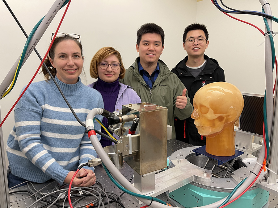
Xu collaborated on the research with UNC-Chapel Hill Professors Christina Inscoe, Jianping Lu and Otto Zhou of the Department of Physics and Astronomy, Don Tyndall of the Adams School of Dentistry and Yueh Lee of the School of Medicine.
The researchers argue that ms-CBCT would be an improvement over conventional CBCT. Conventional CBCT is a specialized medical imaging technique that provides detailed 3-D volumetric images that offer a 360-degree spherical viewing angle of an object. CBCT imaging is used primarily in the fields of dentistry, maxillofacial surgery, interventional radiology and image-guided radiation therapy, where precise visualization of hard-tissue structures like teeth, jaws and the skull is crucial.
“Despite its advantages, conventional CBCT has limitations due to its large imaging volume,” said Xu. “These limitations include reduced soft tissue contrast, image distortion, use that is restricted primarily to imaging hard tissues and hindered quantitative analysis.”
Ms-CBCT overcomes these challenges by enhancing the accuracy of CT imaging of radiodensity of tissues by 60% and soft-tissue contrast by approximately 50%, allowing radiologists and clinicians to distinguish between different tissues and structures within the body. For example, in a CT scan of the abdomen, the liver might have a different HU value than the spleen, helping to identify and characterize abnormalities.
“This is a critical improvement,” said Otto Zhou, David Godschalk Distinguished Professor in the Department of Physics and Astronomy and a member of the APS adjunct faculty, “because accurate HU values are essential for quantitative analysis, tissue characterization and accurate diagnosis.”
In addition to improving image quality and diagnostic accuracy, ms-CBCT reduces the amount of “artifacts,” which can include the presence of metal objects, such as dental fillings or implants, and image distortion due to cone-beam imaging geometry.
“It’s a more comprehensive examination of the imaged area that can be particularly beneficial for capturing larger anatomical structures or regions of interest and improving soft-tissue contrast, making it possible to image more than just hard tissues,” said Xu.
CBCT systems utilize a cone-shaped X-ray beam, as opposed to the fan-shaped beam used in traditional systems. The cone-shaped beam is directed toward the patient or the specific region of interest and a flat-panel detector is positioned opposite the X-ray source. The detector captures the X-rays that pass through the patient, converting them into electrical signals. As the system rotates around the patient, it continuously emits the cone-shaped X-ray beam.
Ms-CBCT involves using multiple X-ray sources configured as an array. Instead of a single, wide cone-angle X-ray tube, this technology employs several X-ray sources that can be strategically positioned around the patient or imaging target. Carbon nanotubes have unique properties that make them suitable for X-ray emission, including their ability to efficiently generate electrons, as well as their small size.
“The ms-CBCT design holds promise for significantly enhancing the capabilities of CBCT scanners,” said Zhou. “These improvements could make CBCT a more versatile tool in medical and dental imaging, potentially competing with multidetector CT images in certain diagnostic applications while retaining its inherent advantages.”
(This article was originally published on the UNC-CH Applied Physical Sciences website. For more details, read the full article.)